AI Applications
Every DeepMathAI application starts as a peer-reviewed publication. We turn our mathematically grounded models into transparent, explainable tools that anyone can use.
Glaucoma Multi-View Screening
AI-powered opportunistic glaucoma screening from retinal fundus images.
Siying, P., et al. & Wangkeeree, R. — Applied Sciences (MDPI), Vol. 16, Issue 7, 3158 (2026)
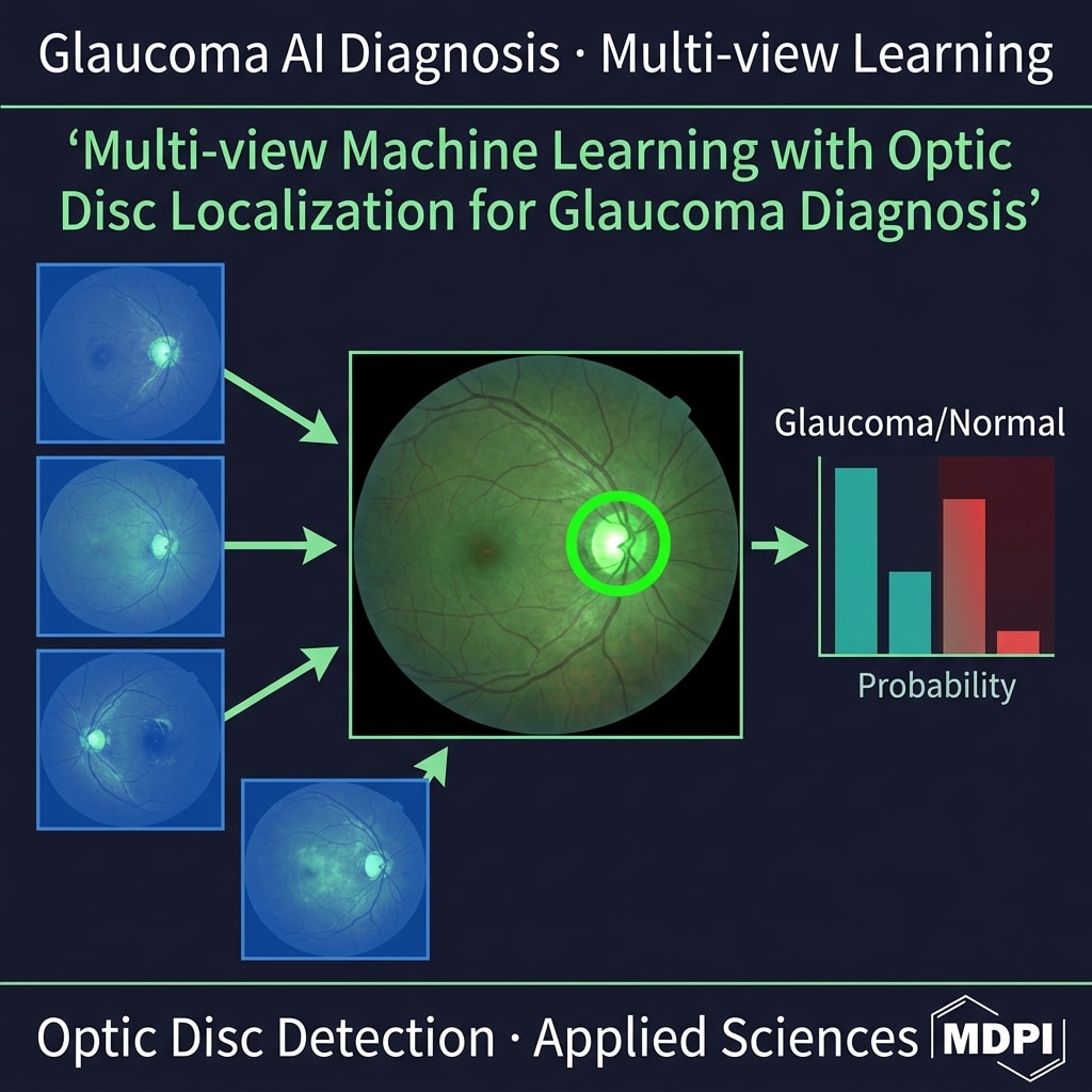
Pipeline
Glaucoma suspected · Grad-CAM ready
How It Works
A web-based decision-support system that flags retinal fundus images as "glaucoma suspected" by fusing predictions from two complementary views of the same image — the full fundus photograph and an automatically localized crop around the optic disc. The multi-view ensemble weights the disc-focused view more heavily, mirroring how ophthalmologists examine the optic nerve head, and every prediction can be inspected through Grad-CAM attribution maps.
Models
- EfficientNetV2-M (full image)
- EfficientNetV2-S (optic-disc crop)
- Weighted multi-view ensemble
Key Features
Single & Batch Prediction
Screen one image or an entire folder of fundus photographs (JPG, PNG, BMP, TIF) in a single run.
Multi-View Fusion
Combines a full-image EfficientNetV2-M and a disc-crop EfficientNetV2-S with weighted probability fusion (Original × 0.5 + Crop × 1.5).
Explainable AI (Grad-CAM)
Heatmaps reveal the retinal regions driving each prediction, supporting clinical trust and error analysis.
Performance Dashboard
Built-in metrics page reporting the model's validated screening performance.
Published Research
Multi-view machine learning with an optic disc localization for glaucoma diagnosis
Siying, P., et al. & Wangkeeree, R. — Applied Sciences (MDPI), Vol. 16, Issue 7, 3158 (2026)
Important limitation: Decision-support tool for screening only — not a medical diagnosis and not a substitute for examination by an ophthalmologist.
VCF MRI2BMD — Osteoporosis Screening
Opportunistic osteoporosis screening from routine lumbar-spine MRI.
Mahatthanatrakul, A., Klinsuwan, T., Wangkeeree, R., & Laoruengthana, A. — Diagnostics (MDPI), Vol. 16, 2241 (2026)
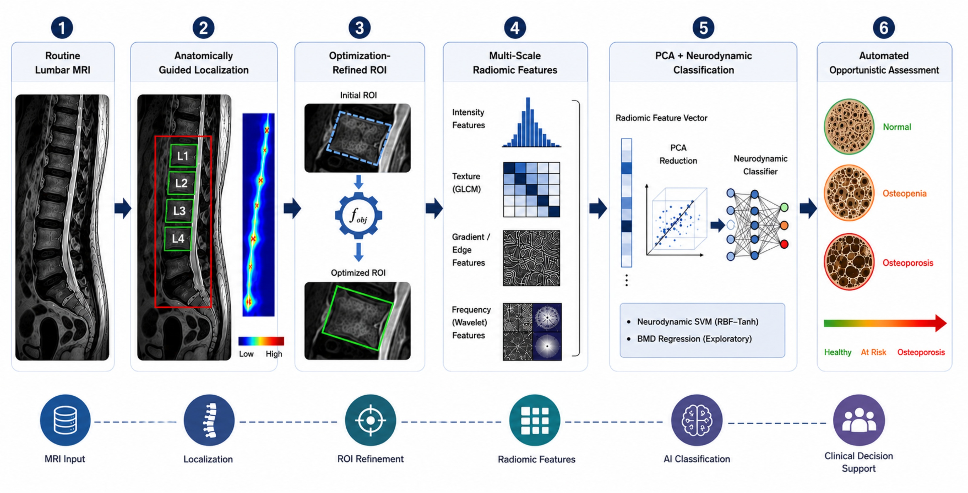
Pipeline
L1–L4 localization → radiomics → BMD assessment
How It Works
Upload a sagittal lumbar-spine MRI slice and the system automatically detects the L1–L4 vertebrae, segments each vertebral body, extracts the study's 546-column radiomic feature vector, and runs it through validated classification and regression models to flag possible osteoporosis and estimate bone mineral density (BMD) and T-score. Because it works on scans acquired for other clinical reasons, it enables early, opportunistic detection at no additional cost to the patient.
Models
- Optimization-refined SVM classifier (BMD status)
- Support Vector Regression for BMD / T-score
- Neurodynamic-SVM (RBF, tanh) — 85.2% published accuracy
Key Features
Anatomically Guided Pipeline
Multi-stage SSIM localization plus mutual-information detection isolates L1–L4, with differential-evolution segmentation of each vertebral body.
546 Radiomic Features
Extracts GLCM, LBP, wavelet, Gabor, Hu-moment, and first-order statistical features per vertebra for a rich, interpretable descriptor.
Single & Batch Prediction
Screen an individual case or queue a batch; each prediction runs as a background job in roughly 10–35 seconds.
Explainable Results
A dedicated explainability view surfaces the features and model reasoning behind each screening decision.
Published Research
An Anatomically Guided and Optimization-Refined Radiomics Framework for Opportunistic Osteoporosis Assessment from Lumbar Spine MRI
Mahatthanatrakul, A., Klinsuwan, T., Wangkeeree, R., & Laoruengthana, A. — Diagnostics (MDPI), Vol. 16, 2241 (2026)
Important limitation: Research decision-support demo — not a diagnostic device. Every output must be confirmed by a radiologist or qualified clinician, and identifiable patient images should never be uploaded.
QMG-RID Explorer — Fundus Image Enhancement
Compare fundus-image enhancement methods live in your browser and watch QMG-RID optimize iteration by iteration.
Wangkeeree, R., Klinsuwan, T., & Luangsawang, K. — manuscript under review (2026)
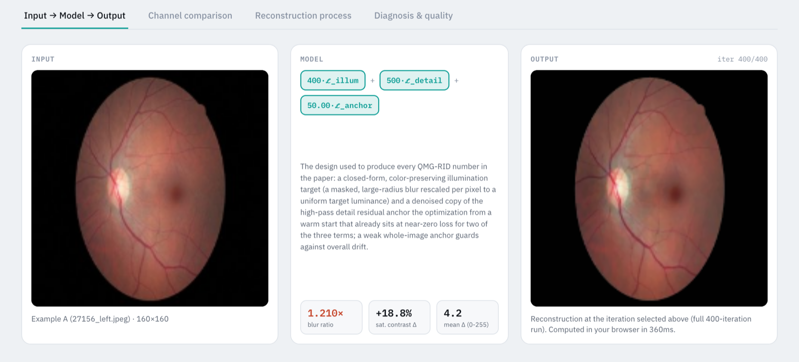
Pipeline
Input → QMG-RID → enhanced output
How It Works
Upload a retinal fundus photograph (or pick an example) and compare enhancement methods side by side: our QMG-RID decomposition, gray-world correction, CLAHE, non-local-means denoising, and the total-variation method of Wang et al. (2021) are all computed live in JavaScript on your device, alongside a precomputed reference from the GAN-based Cofe-Net. QMG-RID splits the image into a base and a detail layer and optimizes them toward a closed-form, color-preserving illumination target and a denoised detail target with per-pixel Adam descent — and the explorer lets you scrub through that optimization step by step.
Models
- QMG-RID — illumination-flattening + detail-preservation decomposition (per-pixel Adam)
- Baselines: gray-world, CLAHE, non-local means, Wang et al. (2021) TV decomposition
- Cofe-Net (Shen et al., 2021) — precomputed reference only
- Frozen DINOv2 ViT-B/14 + 3-class SVM DR classifier — 71.2% 5-fold CV accuracy
Key Features
Runs Entirely In-Browser
Every live method is plain JavaScript running on your own device — uploaded photographs are never sent to a server.
Side-by-Side Method Comparison
Switch between QMG-RID, gray-world, CLAHE, NLM denoising, Wang et al. (2021), and a precomputed Cofe-Net reference, with per-channel (RGB, HSV, CIE L*) before/after views.
Watch the Optimization Unfold
Play or scrub QMG-RID's iterations, inspect its base and detail layers, and toggle each loss term off with ablation presets to see what it contributes.
Diagnosis & Quality Panel
For the example images, real precomputed DR-class predictions and image-quality metrics from the paper's Python pipeline, original vs. enhanced.
Research Manuscript
Image Quality and Enhancement in Diabetic Retinopathy Screening: A Matched-Domain, Cross-Validated Evaluation with Self-Supervised Representations
Wangkeeree, R., Klinsuwan, T., & Luangsawang, K. — manuscript under review (2026)
Important limitation: Research demo accompanying a manuscript under review — not a diagnostic device. The in-browser methods are disclosed, simplified reimplementations at a 160×160 working resolution, so on-screen metrics are illustrative rather than the paper's reported results.
More Applications in Development
Upcoming tools from our research pipeline include diabetic retinopathy screening, vertebral fracture risk stratification, and renewable-energy optimization systems. Follow our publications to see what's next.
Research You Can Actually Use
Interested in collaborating on deployable, explainable AI for healthcare or energy? DeepMathAI welcomes research partnerships and pilot deployments.